CT Scan
A CT (computerized tomography) is also known as a CAT (computer assisted tomography) scan. A CT scan provides cross-sectional X-rays of the body, which are called slices.
CT scans were originally developed for diagnosing disorders of the brain but are now used to image tissues throughout the body. These images can detect tumors, infections and injuries to the internal organs. Bones can be evaluated for fractures and other lesions, and the spinal canal can be assessed for narrowing, nerve impingement and disc herniations. CTA (CT Angiography) is rapidly replacing more invasive catheter studies in evaluation and follow up of vascular disease. The CT provides excellent anatomic detail in the chest, abdomen and pelvis, and many things not visible on traditional X-ray can be seen. Because of the clarity and detail available from a CT scan, patients often avoid exploratory surgery by having a CT scan.
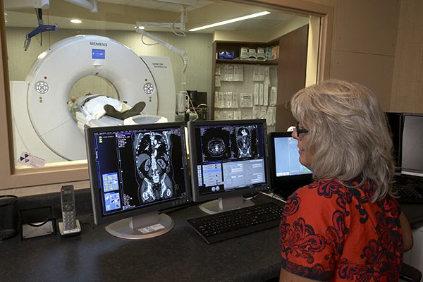
A Greensboro Radiology technologist administers a CT scan
Depending on the area of the body being imaged, patients may be asked to drink a flavored mixture, called contrast. Often, a different type of contrast, or X-ray dye, is injected by a machine through a catheter placed in your vein. Because a CT scan uses an ultra-thin, low dose X-ray beam, radiation exposure is minimized and is often less than that of traditional X-rays.
CT scanners are doughnut-shaped X-ray machines with a flat table through the center. Patients lie on the table as the table moves slowly through the CT scanner. As the table moves, the CT scanner spins around, sending a thin X-ray beam through the body and collecting the emerging beam with a detector. The beam that emerges from the body is collected by the detector, measured by a computer, and converted into a cross-sectional picture, or slice, of your body. Most scans take less than fifteen minutes to complete and some take just a minute or two! Throughout the scan you will be given instructions by the technologist such as when to hold your breath. After a study is done, the images are sent to a special computer system called PACS (Picture Archiving & Communications System) where they are stored and viewed by the radiologist. Patients may be asked to wait until those images are reviewed to ensure additional imaging is not needed. Your doctor can access these images online and review them with you in the office.
*Please tell the doctor ordering a CT and the CT technologist if you are or think you could be pregnant.